

Iron is an essential mineral that plays many important roles in the body. One of its main roles is to transport oxygen via red blood cells throughout the body for energy production. With low iron levels, oxygen saturation is low. Both iron deficiency and overload can cause a host of health problems. Read on if you suspect either.
What is Iron?
Iron (Fe) is an essential element critical for the growth and survival of nearly all organisms [1].
It plays many essential roles in the body. Iron is needed for [2, 3, 4, 5, 6, 7]:
- Red blood cell production
- Oxygen and carbon dioxide transport in the blood (as part of hemoglobin)
- Oxygen transport and storage in muscles (as part of myoglobin)
- Energy production in the heart and muscles
- Brain development and normal brain function
- Immune system development and immune response
- Resistance to infections
- Production and degradation of DNA
- Protecting cells against the accumulation of reactive oxygen species, as a part (cofactor) of enzymes that break down the reactive oxygen species, including oxidases, peroxidases, and catalases
Because of these powerful roles, low iron levels can lead to detrimental effects and eventually, death [8].
However, excessive levels of iron can form reactive oxygen species which lead to tissue and DNA damage [9].
Therefore, it’s important to keep iron levels in balance.
Iron Balance
Iron is one of the most abundant metals in the human body. Most well-nourished adults have approximately 3 – 5 g [10].
Nearly 60% of iron inside the body is incorporated into hemoglobin (in the blood), and 10% into myoglobin (in muscle tissue) [10, 11, 2].
In healthy individuals, the remaining 20 – 30% of iron is stored bound to special proteins, such as transferrin, ferritin, and hemosiderin [10, 11, 2]. These proteins prevent free iron from causing oxidative damage in the body [1, 12, 2, 13, 14].
The liver has the largest capacity to store excess iron [15].
To maintain iron homeostasis, hepcidin, a protein hormone secreted by the liver, inhibits dietary iron absorption in the gut and reduces blood iron levels when iron stores are sufficient [16, 17].
Iron Absorption in the Small Intestine
Cells that line the wall of the small intestine (duodenum and jejunum) help regulate iron absorption based on the iron demands [18, 19].
Heme Iron
Heme iron (iron bound to hemoglobin and myoglobin found in meat) can be directly absorbed by intestinal cells. This process is independent of acidity and is not affected by inhibitors of iron absorption (i.e., phytate and polyphenols) and thus, is more efficient [20].
Non-Heme Iron
Non-heme from plant sources iron is much harder to absorb than heme iron because the intestine can only absorb the ferrous form (Fe2+) and not the ferric form (Fe3+). At physiological (non-acidic) pH, ferrous iron (Fe2+) is readily oxidized to insoluble ferric iron (Fe3+), which cannot be absorbed [21].
Stomach acid helps the enzyme ferric reductase convert Fe3+ into Fe2+, which allows the iron to get absorbed [22]. Therefore, when stomach acid production is impaired (e.g., by acid pump inhibitors), non-heme iron absorption is significantly reduced [23].
Red Blood Cell Recycling
Roughly 1 – 2 mg of dietary iron is absorbed daily, however, processes like hemoglobin synthesis require 20 – 25 mg of iron per day. Most of this iron is obtained through the recycling of old red blood cells by resident macrophages (special white blood cells) [24].
Iron Loss
Around 1 – 2 mg of iron is lost daily through sweat, blood loss, and sloughing of mucosal and skin cells, but there is no organized method of iron excretion in mammals. Therefore, iron levels are balanced by increased (when levels are low) or decreased (when levels are high) absorption of dietary iron in the small intestine [25].
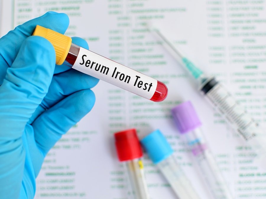
Blood Tests That Evaluate Iron Status
Blood iron tests are typically ordered as follow-up tests when routine tests such as a complete blood count, hemoglobin, and hematocrit levels show abnormal results.
1) Blood Iron
Blood iron measures the amount of circulating iron in the blood. Blood iron is a poor measure of iron status in the body because it fluctuates daily depending on the ingestion of iron-containing foods [26, 27].
A blood iron test without a TIBC or transferrin has limited value except in cases of iron poisoning.
Normal range of iron is around 50-195 mcg/dL or µg/dL (8.95 – 35 µmol/L) in men and 40-190 mcg/dL (7.16 – 34 µmol/L) in women.
Ranges will vary slightly between labs, due to differences in equipment, techniques, and chemicals used.
2) Serum Ferritin
Ferritin levels can serve as a measure of total body iron stores [28].
Low ferritin levels signal that the body’s iron stores are low. Higher levels, on the other hand, may indicate that you have a condition that causes the body to store too much iron [29].
However, ferritin is also an acute phase protein, which means it plays a role in the immune response, and increases in conditions such as chronic inflammation, infections, and cancer, irrespective of iron levels [29, 27, 30].
Read this post to learn more about ferritin.
According to the World Health Organization, the generally accepted cut-off level for blood ferritin levels in which iron stores are depleted is 15 ng/mL for people aged 5 years and older and 12 ng/mL for people younger than 5 years of age [31].
3) Total Iron-Binding Capacity
Total iron-binding capacity (TIBC) measures the total capacity of your blood to bind and transport iron. It is used to estimate the amount of iron stored in your body [32].
TIBC is an indirect measure of transferrin, a protein that binds iron molecules and transports them in the bloodstream [33, 34].
Normal range is around 250 – 450 µg/dL or 44.8 – 76.1 µmol/L. Raised TIBC is characteristic of iron deficiency anemia.
4) Unsaturated Iron-Binding Capacity
UIBC (unsaturated iron-binding capacity) measures the reserve capacity of transferrin, the portion of transferrin that has not yet been saturated with iron. UIBC also reflects transferrin levels.
5) Transferrin Saturation
Transferrin (iron) saturation, also called % saturation, is the percentage of transferrin that is saturated with iron.
Transferrin saturation is calculated by dividing iron levels by total iron-binding capacity (TIBC).
Normally transferrin saturation ranges between 15 – 55 %.
Transferrin saturation <15% indicates iron deficiency, while high levels indicate iron overload (hemochromatosis, transfusional iron overload) [27].
The combined results of transferrin, iron, and TIBC tests are helpful in the differential diagnosis of anemia, iron-deficiency anemia, thalassemia, sideroblastic anemia, and hemochromatosis.
6) Red Cell Zinc Protoporphyrin
When there is an inadequate supply of iron, zinc is incorporated into the protoporphyrin ring of the heme structure, creating zinc protoporphyrin. An elevated zinc protoporphyrin is characteristic of iron-deficient red blood cell production [27].
7) Serum Transferrin Receptor
An elevated serum transferrin receptor (sTfR) is a marker of tissue iron deficiency and increased bone marrow erythropoietic activity.
Since concentrations of transferrin receptor rise when iron stores are depleted to promote cellular iron uptake, they can be used to estimate the magnitude of functional iron deficit once iron stores are depleted [35].
Transferrin levels reflect the extent of red blood cell production and iron demand since the transferrin receptor is mainly derived from developing red blood cells [36].
Advantages of Serum Transferrin Receptor Testing [37, 28, 30]:
- It is an early and sensitive indicator of iron deficiency
- It can distinguish between anemia from chronic disease vs. iron deficiency anemia.
- It is not significantly affected by infection or inflammatory processes, and it does not vary with age, gender, or pregnancy
Normal ranges are between 2.8 – 8.5 mg/L [38].
Low Blood Iron
Iron deficiency is the most common nutritional deficiency in the world, affecting 66 – 80% of the world’s population [39, 40].
It is especially common during pregnancy, affecting 40% – 50% of women and their infants [28, 41].
Iron deficiency is the leading nutritional cause of anemia [39, 40].
Causes
Causes listed below are commonly associated with low iron. Work with your doctor or another health care professional to get an accurate diagnosis. A result that’s lower than normal, doesn’t necessarily mean that you have a health condition needing treatment. Your doctor will interpret your value, taking into account your medical history, symptoms, and other test results, such as hemoglobin, ferritin, transferrin saturation, or TIBC.
1) Insufficient Dietary Intake
Low dietary iron intake can be caused by [42, 43]:
- Malnutrition
- A vegetarian or vegan diet, which lacks heme iron
2) Inadequate Iron Absorption
Several diseases of the digestive system can decrease iron absorption, including [44, 10, 45, 46, 47, 48]:
- Celiac disease
- Irritable bowel diseases, i.e. Crohn’s and ulcerative colitis
- Gastritis
- H. pylori infection
- Small intestinal bacterial overgrowth
- Parasitic worm infections
Bariatric and other weight loss surgeries decrease nutrient absorption, including iron [49].
Other causes of inadequate iron absorption include a high intake of foods or drugs that inhibit iron absorption, are [50, 51]:
- phytates (whole grains, legumes)
- polyphenols (tea, coffee, wine)
- drugs such as antacids, H2 blockers, tetracycline, or cholestyramine [52, 53, 54, 55]
3) Increased Iron Demand
Fast growth increases iron demand, so children, pregnant and lactating women are more likely to be iron deficient [40, 39].
In addition, people who engage in endurance exercise are more likely to have low iron [56].
4) Increased Iron Losses
Bleeding and blood loss increases the loss of iron, including [57, 44, 58]:
- Menstrual bleeding
- Childbirth
- Ulcers
- Hemorrhoids
- Bleeding due to injuries or surgery
- Blood donation
It is important to note that a common and often missed cause of iron deficiency anemia is heavy menstruation. This slow blood loss over time can often result in losing too much iron. Also, because it is a slower process, the body can adapt to the symptoms of iron deficiency and they may be better tolerated. It is important for women with heavy menstruation to have iron levels checked.
Long-term anti-inflammatory drug use (ibuprofen, naproxen, diclofenac) can increase bleeding in the gut.
5) Iron Sequestration
Iron decreases in chronic inflammatory conditions, such as autoimmune disease, chronic infections, chronic kidney disease, or cancer [59, 60, 61, 62, 63]. This is referred to as anemia of chronic disease.
Because iron is important for the growth of pathogens and cancer cells, when there is an infection or inflammation, the body tries to inhibit the growth of pathogens or malignant cells by locking iron away [64].
Tissues affected by infections or inflammation release cytokines that reduce iron blood levels, therefore leading to the development of anemia [65].
Anemia of chronic disease is considered to be a mild-to-moderate form of anemia and treatment is primarily centered on the underlying condition [17].
High-Risk Groups
Groups at higher risk of iron deficiency include:
- Infants and young children [66, 67]
- Obese children [68, 69]
- Women of childbearing age and pregnant women [40, 39]
- Endurance athletes [70]
- Frequent blood donors [44]
- People who have a gut disorder (celiac disease, inflammatory bowel disease, and gut infections) [45]
- People who have had gut surgery (gastrectomy, gut resection, bariatric surgery) [71, 72, 49]
- People with heart failure [73]
- Patients with chronic kidney disease [74]
- Patients with cancer (colon, rectum, stomach; chemotherapy) [60]
Signs and Symptoms
Signs and symptoms of iron deficiency include [75, 72, 76, 77, 78]:
- Fatigue
- Irritability
- Headaches
- Difficulty concentrating
- Pale skin
- Brittle nails
- Hair loss
- Cravings for ice, dirt, clay (pica)
Iron deficiency is associated with lower cognitive test scores, shortened attention span, and reduced physical and mental activity in children and adults [39].
Iron Deficiency Anemia
Iron deficiency anemia (IDA) impacts approximately 1 – 2 billion people worldwide.
In developing countries, it occurs among 23% – 50% of pregnant women and young children [41].
IDA is characterized by a defect in hemoglobin synthesis, resulting in the reduced capacity of the red blood cells to deliver oxygen to tissues [79].
IDA can become severe, and cause lethargy, pale skin, shortness of breath, irritability, decreased appetite, failure to thrive, and heart failure [80, 81].
Children and women are at higher risk. Iron deficiency can result in preterm birth, poor growth and cognitive skills, and neurological dysfunction [75].
Iron deficiency represents a spectrum ranging from iron depletion, which causes no biological impairments, to iron-deficiency anemia, which affects the functioning of several organ systems [82].
Iron deficiency can be divided into 3 stages, including [83]:
Phase 1: Pre-latent Stage – Depleted Iron Stores
Iron stores are lowered or absent, serum iron concentration, hemoglobin, and hematocrit are normal. This stage of iron deficiency is manifested with the reduction or absence of bone marrow iron stores and reduced serum ferritin levels.
Phase 2: Latent Stage
Serum iron (SI) and transferrin saturation are reduced in addition to reduced iron stores. Hemoglobin and hematocrit are still within normal limits.
Phase 3: Iron Deficiency Anemia
During this phase, in addition to the depletion of iron stores, serum iron, and transferrin saturation, hemoglobin and hematocrit levels are reduced.
The red blood cells of individuals that have iron-deficiency anemia are smaller and paler than normal [84].
Genetics of Iron Deficiency
Iron-refractory iron deficiency anemia is caused by a rare mutation in a gene (encoding Matriptase-2, an iron regulatory enzyme) expressed in the liver, which leads to high hepcidin levels. As a result, iron absorption from the intestine and release from macrophages is inhibited, resulting in severe iron deficiency [85, 86].
Genetic Variants that are Associated with Anemia (SelfDecode)
| Genes | SNP | Risk Alleles |
| HFE | rs1800562 | A, A |
| LOC100507006, LOC105374768 | rs2698530 | C, C, C |
| TF | rs3811647 | A, A |
| IGLV4-60, LOC102724653 | rs987710 | G, G |
High Blood Iron
Causes
Causes listed below are commonly associated with high iron. Work with your doctor or another health care professional to get an accurate diagnosis. A result that’s higher than normal, doesn’t necessarily mean that you have a health condition needing treatment. Your doctor will interpret your value, taking into account your medical history, symptoms, and other test results, such as hemoglobin, ferritin, transferrin saturation, or TIBC.
1) Acute Iron Poisoning
Iron toxicity (acute iron overload) occurs mainly in children. Taking 20 mg of elemental iron per kg of body weight causes vomiting and diarrhea. In severe cases, taking about 60 mg per kg body weight (11 of commonly sold 27 mg tablets of ferrous sulfate) causes blood loss, multiple organ dysfunction, and death [87, 88, 89].
2) Chronic Iron Overload
Chronic iron overload can be caused by:
- Repeated blood transfusions [90]
- Iron loading anemias: thalassemia major, sideroblastic anemia, aplastic anemia [90, 91]
- Alcoholism (increased uptake of iron) [92, 93, 94]
- Liver scarring (cirrhosis) and liver disease [95, 96, 97, 98]
- Hereditary hemochromatosis, an inherited disorder in which your body absorbs too much iron from the food that you eat [99, 91]
- Conditions that cause abnormal destruction of red blood cells (hemolysis) [100]
- Chloramphenicol, an antibiotic used to treat bacterial infections [101, 102]
The body’s rate of iron absorption depends on the action of hepcidin, a circulating hormone secreted by the liver that decreases iron absorption. Most hereditary iron disorders like hemochromatosis result from mutations in genes affecting hepcidin production [103, 104].